Nature Communications: Slim, a new slide microscopy viewing tool
Nature Communications: Slim, an open-source, web-based slide microscopy tool
New viewer and annotation tool closes the gap between radiology and pathology imaging
A team of researchers led by Markus Herrmann, MD, PhD, Director of Computational Pathology at Massachusetts General Hospital and Assistant Professor of Pathology at Harvard Medical School, have developed Slim, an open-source, web-based slide microscopy viewer and annotation tool that closes the gap between radiology and pathology imaging to facilitate integrated, multi-modal imaging data science, to drive interdisciplinary cancer research across departmental boundaries.
Their paper about the development of this tool was published in Nature Communications, in March 2023: Interoperable slide microscopy viewer and annotation tool for imaging data science and computational pathology
As the authors note, cancer researchers increasingly use machine learning (ML) to derive insights from biomedical images and require access to large amounts of imaging data to train and validate ML models. The NCI Imaging Data Commons (IDC) is a cloud platform that hosts large public collections of cancer imaging data from multiple research projects and institutions across a wide range of modalities and domains.
IDC provides web-based interfaces and tools to query, visualize, annotate, and analyze the data, thereby enabling cancer researchers to build cohorts for ML model development using diverse data. Data standardization is essential in aggregating diverse imaging datasets and in making the data findable, accessible, interoperable, and reusable (FAIR). The IDC has harmonized all data using the internationally accepted Digital Imaging and Communication in Medicine (DICOM) standard. While DICOM is well established in radiology and many open-source tools exist to visualize DICOM data in research setting, DICOM adoption in pathology is pending and DICOM-based tools are lacking.
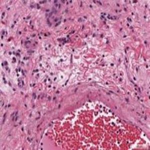
The new tool – Slim – serves as the microscopy viewer of the IDC, where it enables researchers to visualize brightfield as well as highly multiplexed immunofluorescence microscopy images alongside a wealth of other clinical cancer imaging data, including computed tomography and positron emission tomography images.
The team that developed this new slide microscopy viewer used imaging data housed in the IDC, from various NCI-funded programs:
• The Cancer Genome Atlas (TCGA)
• Human Tumor Atlas Network (HTAN)
• Clinical Proteomic Tumor Analysis Consortium (CPTAC)
Using Slim, they also demonstrate how to collect user-drawn image annotations essential for ML model development in DICOM format, as well as how to visualize computational image analysis results and ML model outputs in DICOM format, including image segmentation masks, heatmaps, and single-cell contours.
They further demonstrate the ability of Slim to interoperate with various commercial digital pathology systems to streamline the translation of novel image-based methods and tools from research settings into clinical practice.
“Aggregating large and complex slide microscopy imaging data from various biomedical research and clinical labs and integrating data from multiple modalities across medical domains is a major challenge for computational pathology and cancer imaging research, but it also represents an immense, and largely untapped opportunity for new discoveries and innovations for the benefit of cancer patients,” said Dr. Hermann. “Massive scale and different modalities are required to more rapidly advance our understanding of how various cancer types and stages manifest morphologically across different length scales and over time, and to develop novel computational image analysis solutions and quantitative imaging biomarkers for improving cancer care and outcomes.”
By relying on the open-source web-based DICOM application, the Slim tool can be used by any browser, drawing from cloud-based or on-prem datasets, and can standardize pixel level image information including colors and luminosity/pixel intensity.
“The standardization of images from various research and clinical imaging platforms that are developed by a range of commercial vendors, has been a complicating factor in the aggregation of imaging data across large research projects and consortia,” said Andrey Fedorov, PhD, principle investigator at the Surgical Planning Laboratory at Brigham and Women’s Hospital and Associate Professor of Radiology at Harvard Medical School, who leads the development of the IDC together with Ron Kikinis, MD, Professor of Radiology at Harvard Medical School. “Slim demonstrates that the same DICOM standard we have been using for decades in radiology applications can support digital pathology and microscopy, allowing us to leverage standard server-side interfaces for interacting with the data harmonized into DICOM across domains, vendors and modalities.”
IDC is a cloud-based repository of publicly available cancer imaging data co-located with the analysis and exploration tools and resources. IDC is a node within the broader NCI Cancer Research Data Commons (CRDC) infrastructure that provides secure access to a large, comprehensive, and expanding collection of cancer research data.
Learn more about the Imaging Data Commons (IDC).